37 of 37 spec points in this unit have notes.
- Notes
- Suggested skills activity
- Coming soon
-
Eukaryotic Cell Structure
Spec points
Organelle Structure & FunctionJump to notes →
The structure of eukaryotic cells, restricted to the structure and function of:
• cell-surface membrane
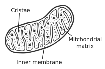
• nucleus (containing chromosomes, consisting of protein-bound, linear DNA, and one or more nucleoli)
• mitochondria
• chloroplasts (in plants and algae)
• Golgi apparatus and Golgi vesicles
• lysosomes (a membrane-bound organelle that releases hydrolytic enzymes)
• ribosomes
• rough endoplasmic reticulum and smooth endoplasmic reticulum
• cell wall (in plants, algae and fungi)
• cell vacuole (in plants).Cell Specialisation & OrganisationJump to notes →
In complex multicellular organisms, eukaryotic cells become specialised for specific functions. Specialised cells are organised into tissues, tissues into organs and organs into systems.Explaining Cell AdaptationsJump to notes →
Students should be able to apply their knowledge of these features in explaining adaptations of eukaryotic cells.
-
Prokaryotic Cells & Viruses
Spec points
Prokaryotic Cell StructureJump to notes →
Prokaryotic cells are much smaller than eukaryotic cells. They also differ from eukaryotic cells in having:
• cytoplasm that lacks membrane-bound organelles
• smaller ribosomes
• no nucleus; instead they have a single circular DNA molecule that is free in the cytoplasm and is not associated with proteins
• a cell wall that contains murein, a glycoprotein.
In addition, many prokaryotic cells have:
• one or more plasmids
• a capsule surrounding the cell
• one or more flagella.
Details of these structural differences are not required.Structure of VirusesJump to notes →
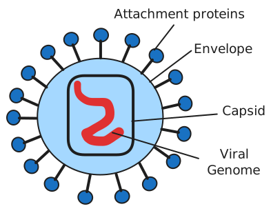
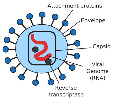
Viruses are acellular and non-living. The structure of virus particles to include genetic material, capsid and attachment protein.
-
Methods of Studying Cells
Spec points
Principles & Limitations of MicroscopesJump to notes →
The principles and limitations of optical microscopes, transmission electron microscopes and scanning electron microscopes.Magnification & ResolutionMS 1.8Jump to notes →
Measuring the size of an object viewed with an optical microscope. The difference between magnification and resolution.
Use of the formula: magnification = size of image / size of real objectCell Fractionation & UltracentrifugationJump to notes →
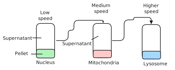
Principles of cell fractionation and ultracentrifugation as used to separate cell components.Distinguishing Artefacts from OrganellesJump to notes →
Students should be able to appreciate that there was a considerable period of time during which the scientific community distinguished between artefacts and cell organelles.Identifying Starch Grains in Plant CellsSkillAT dAT eAT fJump to notes →
Students could use iodine in potassium iodide solution to identify starch grains in plant cells.
-
All Cells Arise from Other Cells
Spec points
The Cell CycleJump to notes →
Within multicellular organisms, not all cells retain the ability to divide.
Eukaryotic cells that do retain the ability to divide show a cell cycle.
• DNA replication occurs during the interphase of the cell cycle.
• Mitosis is the part of the cell cycle in which a eukaryotic cell divides to produce two daughter cells, each with the identical copies of DNA produced by the parent cell during DNA replication.Stages of MitosisJump to notes →
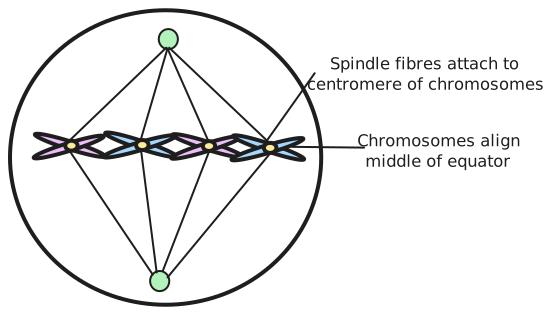
The behaviour of chromosomes during interphase, prophase, metaphase, anaphase and telophase of mitosis. The role of spindle fibres attached to centromeres in the separation of chromatids.
Division of the cytoplasm (cytokinesis) usually occurs, producing two new cells.
Students should be able to:
• recognise the stages of the cell cycle: interphase, prophase, metaphase, anaphase and telophase (including cytokinesis)
• explain the appearance of cells in each stage of mitosis.Uncontrolled Cell Division: Tumours & CancerJump to notes →
Mitosis is a controlled process. Uncontrolled cell division can lead to the formation of tumours and of cancers. Many cancer treatments are directed at controlling the rate of cell division.Binary Fission in ProkaryotesJump to notes →
Binary fission in prokaryotic cells involves:
• replication of the circular DNA and of plasmids
• division of the cytoplasm to produce two daughter cells, each with a single copy of the circular DNA and a variable number of copies of plasmids.Viral ReplicationJump to notes →
Being non-living, viruses do not undergo cell division. Following injection of their nucleic acid, the infected host cell replicates the virus particles.Required Practical 2Required practicalAT dAT eMS 1.8MS 0.3Jump to notes →
Required practical 2: Preparation of stained squashes of cells from plant root tips; set-up and use of an optical microscope to identify the stages of mitosis in these stained squashes and calculation of a mitotic index.
Students should measure the apparent size of cells in the root tip and calculate their actual size using the formula:
Actual size = size of image / magnification
Calculation of a mitotic index.
-
Transport Across Cell Membranes
Spec points
Membrane Structure & the Fluid-Mosaic ModelJump to notes →
The basic structure of all cell membranes, including cell-surface membranes and the membranes around the cell organelles of eukaryotes, is the same.
The arrangement and any movement of phospholipids, proteins, glycoproteins and glycolipids in the fluid-mosaic model of membrane structure. Cholesterol may also be present in cell membranes where it restricts the movement of other molecules making up the membrane.Simple DiffusionJump to notes →
• simple diffusion (involving limitations imposed by the nature of the phospholipid bilayer)Facilitated DiffusionJump to notes →
• facilitated diffusion (involving the roles of carrier proteins and channel proteins)OsmosisJump to notes →
• osmosis (explained in terms of water potential)Active TransportJump to notes →
• active transport (involving the role of carrier proteins and the importance of the hydrolysis of ATP)Co-TransportJump to notes →
• co-transport (illustrated by the absorption of sodium ions and glucose by cells lining the mammalian ileum).Adaptations & Factors Affecting Rate of TransportJump to notes →
Cells may be adapted for rapid transport across their internal or external membranes by an increase in surface area of, or by an increase in the number of protein channels and carrier molecules in, their membranes.
Students should be able to:
• explain the adaptations of specialised cells in relation to the rate of transport across their internal and external membranes
• explain how surface area, number of channel or carrier proteins and differences in gradients of concentration or water potential affect the rate of movement across cell membranes.Required Practical 3Required practicalMS 3.2MS 3.4Jump to notes →
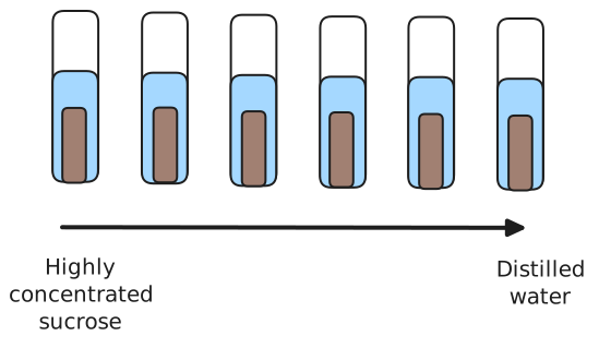
Required practical 3: Production of a dilution series of a solute to produce a calibration curve with which to identify the water potential of plant tissue.
Students could plot the data from their investigations in an appropriate format.
Students could determine the water potential of plant tissues using the intercept of a graph of, eg, water potential of solution against gain/loss of mass.Required Practical 4Required practicalMS 3.2Jump to notes →
Required practical 4: Investigation into the effect of a named variable on the permeability of cell-surface membranes.
Students could plot the data from their investigations in an appropriate format.
-
Cell Recognition & Immune System
Spec points
Cell Recognition & AntigensJump to notes →
Each type of cell has specific molecules on its surface that identify it. These molecules include proteins and enable the immune system to identify:
• pathogens
• cells from other organisms of the same species
• abnormal body cells
• toxins.
Definition of antigen. The effect of antigen variability on disease and disease prevention.PhagocytosisJump to notes →
Phagocytosis of pathogens. The subsequent destruction of ingested pathogens by lysozymes.Cellular Response: T LymphocytesJump to notes →
The response of T lymphocytes to a foreign antigen (the cellular response).
• The role of antigen-presenting cells in the cellular response.
• The role of helper T cells (T_H cells) in stimulating cytotoxic T cells (T_C cells), B cells and phagocytes. The role of other T cells is not required.Humoral Response: B LymphocytesJump to notes →
The response of B lymphocytes to a foreign antigen, clonal selection and the release of monoclonal antibodies (the humoral response).Antibody Structure & ActionJump to notes →
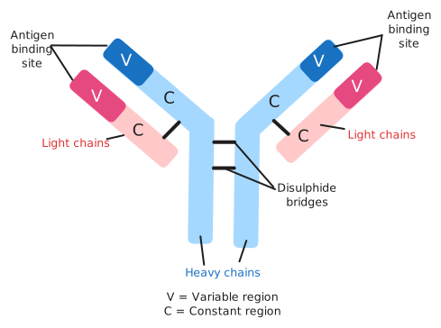
• Definition of antibody.
• Antibody structure.
• The formation of an antigen-antibody complex, leading to the destruction of the antigen, limited to agglutination and phagocytosis of bacterial cells.Primary & Secondary Immune ResponsesJump to notes →
• The roles of plasma cells and of memory cells in producing primary and secondary immune responses.Vaccination & Herd ImmunityJump to notes →
The use of vaccines to provide protection for individuals and populations against disease. The concept of herd immunity.Active & Passive ImmunityJump to notes →
The differences between active and passive immunity.
-
HIV and AIDS
Spec points
HIV Structure & ReplicationJump to notes →
Structure of the human immunodeficiency virus (HIV) and its replication in helper T cells.AIDS & Why Antibiotics Don't Work on VirusesJump to notes →
How HIV causes the symptoms of AIDS. Why antibiotics are ineffective against viruses.
-
Monoclonal Antibodies & ELISA
Spec points
Ethical Issues & Evaluating EvidenceJump to notes →
Ethical issues associated with the use of vaccines and monoclonal antibodies.
Students should be able to:
• discuss ethical issues associated with the use of vaccines and monoclonal antibodies
• evaluate methodology, evidence and data relating to the use of vaccines and monoclonal antibodies.Uses of Monoclonal AntibodiesJump to notes →
The use of monoclonal antibodies in:
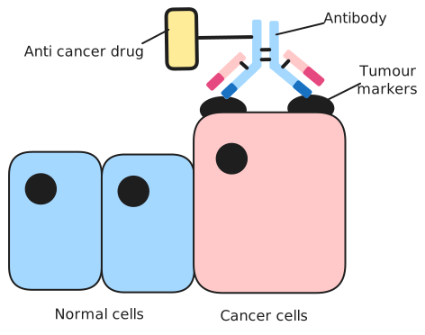
• targeting medication to specific cell types by attaching a therapeutic drug to an antibody
• medical diagnosis.
Details of the commercial or scientific production of monoclonal antibodies are not required.The ELISA TestJump to notes →
The use of antibodies in the ELISA test.