Gene Mutations
What you need to know (based on the AQA specification)
What you need to know (based on the AQA specification)
Gene mutations involve a change in the base sequence of chromosomes. They can arise spontaneously during DNA replication and include base deletion and base substitution. Due to the degenerate nature of the genetic code, not all base substitutions cause a change in the sequence of encoded amino acids. Mutagenic agents can increase the rate of gene mutation.
A gene mutation is a change in the base sequence of chromosomal DNA. Every mutation creates a new allele of the gene.
Gene mutations can occur spontaneously during DNA replication, or can be triggered by mutagenic agents — factors that increase the rate of mutation:
- UV radiation
- Ionising radiation (e.g. X-rays, gamma rays)
- Chemical mutagens (e.g. some components of cigarette smoke)
Types of Gene Mutation
There are two main types you need to know (for Unit 4, there are additional in Unit 8).
Base Substitution
One base is replaced by a different base (e.g. A replaced by G). This changes a single codon. Depending on what the new codon codes for, the effect varies:
- Silent — the new codon codes for the same amino acid (because the genetic code is degenerate). No change in the polypeptide.
- Missense — the new codon codes for a different amino acid, potentially altering the protein’s tertiary structure and function.
- Nonsense — the new codon is a stop codon, ending translation early and producing a truncated, non-functional polypeptide.
Base Deletion (and Addition)
A base is removed (deletion) or added (addition/insertion). Because codons are read in groups of three, this shifts the reading frame of every codon downstream. This causes a frameshift mutation, altering every amino acid from that point onwards.
Why is a frameshift usually so much worse than a substitution?
Why is a frameshift usually so much worse than a substitution?
A substitution changes one codon at most. One amino acid is affected. A frameshift shifts the reading frame for every codon downstream. That means potentially every amino acid from the mutation site onwards is wrong, almost certainly producing a non-functional protein.
Why doesn't a mutation always change the amino acid sequence?
Why doesn't a mutation always change the amino acid sequence?
The genetic code is degenerate: most amino acids are coded for by more than one codon. A base change often still codes for the same amino acid. Also, if the mutation is in a non-coding region (an intron or between genes), it won’t affect the polypeptide at all.
Common exam mistake — mutation chain of reasoning
When explaining how a mutation affects a protein, follow the full chain: changed base sequence → changed codon → changed amino acid → changed primary structure → changed tertiary structure → changed function. Many students stop too early, e.g. saying “the protein changes shape” without linking back to the amino acid sequence change.
You can explore specific mutation examples in gene mutations in Unit 8.
Chromosome Mutations
What you need to know (based on the AQA specification)
What you need to know (based on the AQA specification)
Mutations in the number of chromosomes can arise spontaneously by chromosome non-disjunction during meiosis.
So far we’ve looked at changes to the DNA base sequence. But mutations can also affect whole chromosomes.
Non-disjunction occurs when homologous chromosomes (meiosis I) or sister chromatids (meiosis II) fail to separate properly. This produces gametes with an abnormal chromosome number:
- Some gametes receive an extra chromosome (n + 1)
- Some gametes receive one fewer chromosome (n − 1)
If this gamete is then fertilised by a normal gamete, the resulting zygote has an abnormal chromosome number. For example, non-disjunction of chromosome 21 can lead to trisomy 21 (Down syndrome).
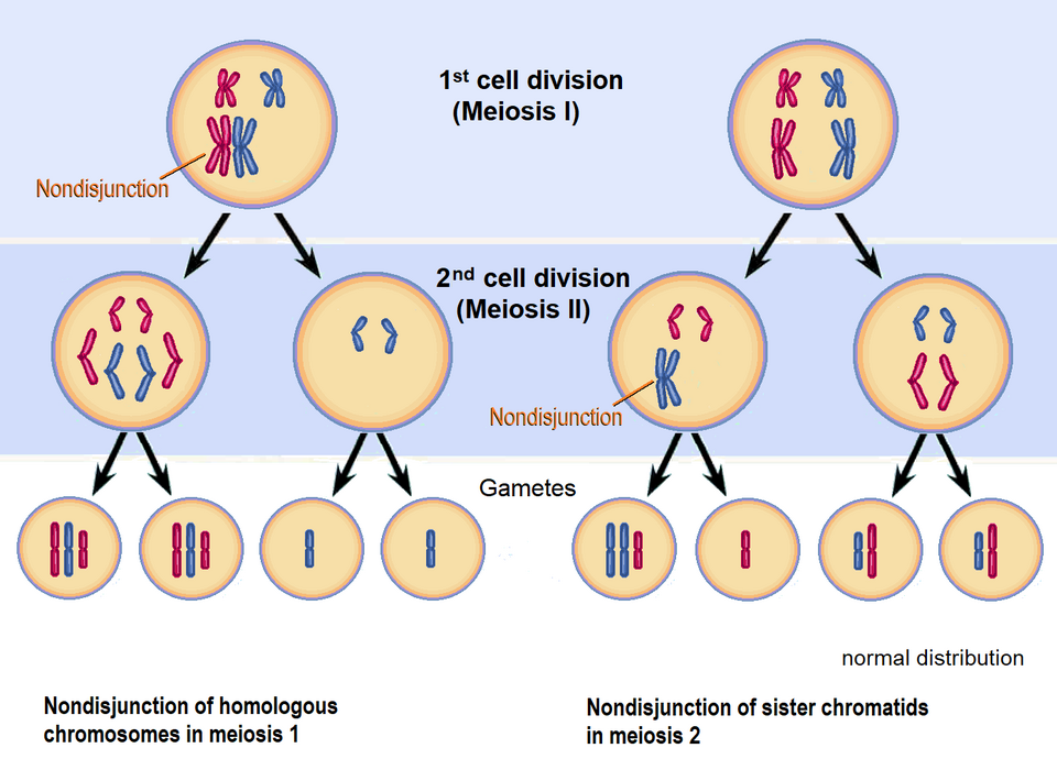
Image: Wikimedia Commons, CC BY-SA 3.0
Common exam mistake — non-disjunction
Non-disjunction affects one chromosome pair only, not all chromosomes. A gamete produced by non-disjunction still carries almost all chromosomes correctly; only the one affected pair is abnormal (n+1 or n−1).
Meiosis
Important - What you need to know about meiosis
Important - What you need to know about meiosis
Meiosis produces daughter cells that are genetically different from each other.
The process of meiosis in sufficient detail to show how:
- two nuclear divisions result usually in the formation of four haploid daughter cells from a single diploid parent cell
- genetically different daughter cells result from the independent segregation of homologous chromosomes
- crossing over between homologous chromosomes results in further genetic variation among daughter cells.
Why is meiosis needed?
When a sperm and egg fuse at fertilisation, each contributes half the genetic information, so meiosis halves the chromosome number (2n → n) in advance, ensuring the resulting zygote has the correct diploid number (2n).
Meiosis involves two successive divisions, producing four haploid cells that are genetically non-identical to each other.
Before meiosis begins, the cell must complete interphase: DNA replication occurs and the cell grows, ensuring each daughter cell will receive a complete set of chromosomes.
What are homologous chromosomes?
What are homologous chromosomes?
A matching pair of chromosomes — one inherited from mum, one from dad — that carry the same genes at the same positions (loci), but may carry different alleles of those genes.
| Homologous chromosomes | Sister chromatids | |
|---|---|---|
| Origin | One maternal, one paternal | Identical copies from DNA replication |
| Genetic content | Same genes, possibly different alleles | Genetically identical |
| Joined at centromere? | No | Yes |
| Separate in… | Meiosis I | Meiosis II / Mitosis |
In a diploid human cell there are 23 pairs of homologous chromosomes (46 total).
Meiosis I
In meiosis I, the homologous chromosome pairs separate and it reduces the chromosome number by half.

- Prophase I — Homologous chromosomes pair up to form bivalents. Non-sister chromatids may exchange sections of DNA at chiasmata — this is crossing over
- Metaphase I — Homologous pairs of chromosomes line up randomly at the equator (centre of the cell) - this is random (independent segregation)
- Anaphase I — Homologous chromosomes are pulled to opposite poles; chromatids remain joined at their centromeres
- Telophase I / Cytokinesis — Two haploid cells form, each containing one chromosome from each homologous pair (still as two chromatids)
Meiosis II
In meiosis II, the sister chromatids separate, similar in mechanism to mitosis.

- Chromosomes align at the equator of each cell (metaphase 2)
- Centromeres split and chromatids are pulled to opposite poles (anaphase 2)
- Four haploid daughter cells are produced, genetically non-identical to each other
Meiosis vs Mitosis
| Meiosis | Mitosis | |
|---|---|---|
| Divisions | 2 | 1 |
| Daughter cells | 4 | 2 |
| Ploidy | Haploid (n) | Diploid (2n) |
| Genetic identity | Non-identical | Identical |
| Purpose | Gamete formation | Growth / repair |
Sources of Genetic Variation
Meiosis creates genetic diversity in two ways during meiosis I, with random fertilisation adding further variation afterwards.
Crossing Over
During prophase I, non-sister chromatids of homologous chromosomes break and rejoin at chiasmata, exchanging sections of DNA. This produces recombinant chromosomes with new combinations of alleles that weren’t present in either parent chromosome.

Independent Segregation
During metaphase I, homologous chromosome pairs line up randomly at the equator and independently of every other pair (Independently means that one pair does not affect the other pair). So each gamete gets a different random mix of maternal and paternal chromosomes - there are different combinations

Random Fertilisation
Any male gamete can fuse with any female gamete. This random fusion further increases genetic variation in offspring.
Calculating different combinations
The number of possible chromosome combinations from independent assortment is 2ⁿ, where n = the number of homologous pairs.
For humans (n = 23): 2²³ ≈ 8 million possible gametes from independent assortment alone.
When a sperm and egg fuse, each gamete independently carries 2²³ combinations, so the number of possible offspring is:
2²³ × 2²³ = 2⁴⁶ ≈ 70 trillion
And this is before crossing over is even considered, which increases variation further still.
Sources of genetic variation — summary
The following processes produce new combinations of alleles:
- Crossing over between homologous chromosomes (Prophase I)
- Independent segregation of homologous chromosomes (Metaphase I)
- Random fertilisation of gametes
The non-coding repeats between genes that arise from this variation are also the basis of DNA profiling / genetic fingerprinting in Unit 8.
Exam Questions
Define ‘gene mutation’ and explain how a gene mutation can have:
- no effect on an individual
- a positive effect on an individual
Hint
For the definition: what changes, and what does this create? For ‘no effect’: think about the degenerate code AND non-coding DNA. For ‘positive effect’: think survival and reproductive success.
Mark Scheme
- (Definition) Change in the base / nucleotide sequence of chromosomal DNA (1 mark)
- Results in the formation of a new allele (1 mark)
- (No effect because) Genetic code is degenerate so amino acid sequence may not change (1 mark) OR Mutation is in an intron so amino acid sequence may not change (1 mark)
- (Positive effect because) Results in change in polypeptide that positively changes the properties of the protein (1 mark) May result in increased reproductive success / increased survival (1 mark)
(For full marks: at least one mark must be scored in each section)
Tips from examiner reports
- A gene mutation does NOT always change the amino acid sequence — the degenerate code is the key explanation
- Any mutation creates a new allele — state this explicitly
- For a beneficial effect, the key idea is increased survival and reproductive success — not just ‘changed appearance’
Mutation can result in an increase in genetic variation within a species. Describe and explain the other processes (apart from mutation) that result in increases in genetic variation.
(4 marks)Hint
Focus on meiosis: crossing over, independent segregation, and fertilisation. How does each process create NEW COMBINATIONS of alleles?
Mark Scheme
- Independent segregation of homologous chromosomes/pairs (1 mark)
- Crossing over between homologous chromosomes/pairs (1 mark)
- Random fertilisation of gametes (1 mark)
- (These processes produce) new combinations of alleles (1 mark)
Comments from mark scheme
- For ‘independent’ accept ‘random’
- For ‘segregation’ accept ‘assortment’
- For crossing over: accept ‘within bivalent’ for ‘between homologous pair’
- Accept an additional mark: ‘produces new combinations of maternal and paternal chromosomes’
- Ignore reference to epigenetics
Tips from examiner reports
- For genetic variation in meiosis, include: crossing over (between homologous chromosomes during prophase I), independent assortment/segregation, and random fertilisation
- Don’t forget to mention homologous chromosome pairing before describing crossing over
- Random fertilisation involves gametes — say so explicitly
- Migration introduces new alleles but doesn’t create new allele combinations
Complete Figure 6 to show the chromosomes inside the daughter cells formed after the second meiotic division.
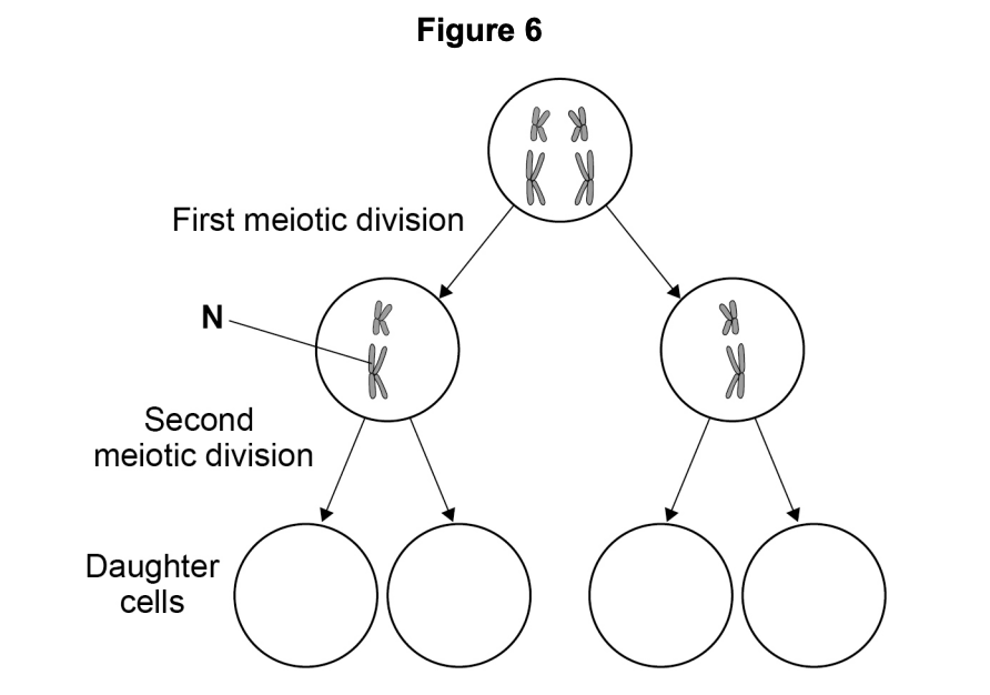
Hint
The right-hand cell undergoes normal Meiosis II: sister chromatids separate as usual. The left-hand cell has non-disjunction of chromosome N. Both chromatids of N go to the same pole.
Mark Scheme
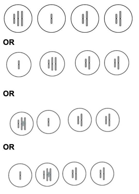
- 2 cells on left correct — one cell receives both chromatids of chromosome N (n+1), the other receives none (n−1), both also carry the other chromosome (1 mark)
- 2 cells on right correct — normal separation, each receives one chromatid of each chromosome (1 mark)
(Ignore differences in chromosome length in drawn cells)
Comments from mark scheme
- Accept any valid arrangement showing the non-disjunction effect on the left pair
- Ignore minor drawing differences in chromosome length
Tips from examiner reports
- Non-disjunction means chromatids fail to separate — one cell gets both, one gets none
- Non-disjunction of one pair doesn’t affect the other chromosome — only the N pair is abnormal
- Draw chromatids clearly — show the difference between long and short chromosomes
Comments from mark scheme