What you need to know (based on the AQA specification)
What you need to know (based on the AQA specification)
- The use of monoclonal antibodies in:
- targeting medication to specific cell types by attaching a therapeutic drug to an antibody
- medical diagnosis.
- The use of antibodies in the ELISA test.
- Ethical issues associated with the use of vaccines and monoclonal antibodies.
Details of the commercial or scientific production of monoclonal antibodies are not required.
Monoclonal antibodies are identical antibodies produced from a single clone of plasma cells — so they all bind to the same specific antigen. Their specificity makes them powerful tools in medicine (targeted treatment) and diagnosis (e.g. ELISA, pregnancy tests).
Uses of Monoclonal Antibodies
Targeting Medication to Specific Cell Types
Because monoclonal antibodies bind to one specific antigen, a therapeutic drug can be attached to the antibody, which delivers the drug only to cells with that antigen.
Example — treating cancer:
- Cancer cells have antigens on their surface that aren’t found on healthy cells — tumour markers
- Monoclonal antibodies are made that are complementary to the tumour marker
- An anti-cancer drug is attached to the antibody
- The antibodies are injected into the bloodstream — they bind only to the cancer cells
- The drug enters and destroys the cancer cells, without affecting healthy cells

This means:
- Smaller doses of drug are needed (cheaper, fewer side effects)
- Side effects are reduced (drug doesn’t affect healthy cells)
Medical Diagnosis
Monoclonal antibodies can be used to detect specific molecules in the body, useful for diagnosis.
Example — pregnancy tests:
- During pregnancy, the hormone hCG (human chorionic gonadotropin) is produced and appears in urine
- The pregnancy test contains monoclonal antibodies complementary to hCG, attached to a coloured dye
- If hCG is present in the urine, it binds to the antibodies, and a coloured line appears

The ELISA Test
ELISA (Enzyme-Linked Immunosorbent Assay) is a test that uses antibodies to detect the presence (and amount) of a specific antigen or antibody in a sample.
It works by using an enzyme-linked antibody to bind the target molecule. Once bound, a substrate is added. The enzyme converts it into a coloured product, giving a visible positive result.
There are two versions:
- Direct ELISA — the enzyme-linked antibody binds the target antigen directly
- Indirect ELISA — a primary antibody binds the target first; then a secondary, enzyme-linked antibody binds the primary antibody (this is the version used in the HIV test below)

Example — testing for HIV antibodies
- HIV antigens are bound to the inside of a well in a plate
- The patient’s blood sample is added — any HIV antibodies in their blood bind to the HIV antigens
- The well is washed to remove unbound antibodies
- A secondary antibody (with an enzyme attached) is added — it binds to the patient’s HIV antibodies
- The well is washed again to remove unbound secondary antibodies
- A substrate for the enzyme is added — the enzyme converts it into a coloured product
- If a colour appears, the patient has HIV antibodies — they have been exposed to HIV
The intensity of the colour can be measured with a colorimeter to quantify how much antibody is present.
The same method can be used to detect an antigen instead (e.g. a pathogen). Antibodies are bound to the well, and the antigen in the sample binds them.
Why are the wash steps so important?
Why are the wash steps so important?
The washing steps remove unbound antibodies. If you didn’t wash, any antibody could give a colour change — even ones that didn’t actually bind — giving a false positive.
Ethical Issues
Answering 'evaluate the ethics' questions
Always give both sides:
- One concern (e.g. animal testing, drug-trial risks, cost / unequal access)
- One benefit (e.g. saves lives, prevents disease, effective treatment)
Exam Question Practice
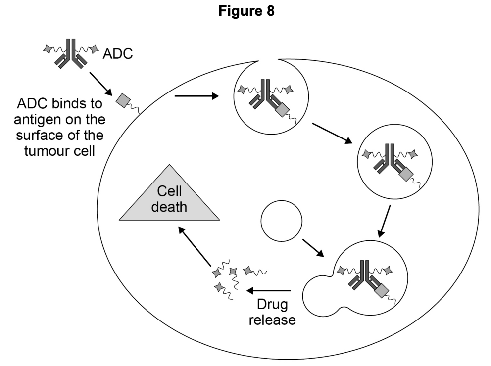
ADCs are molecules made of a monoclonal antibody linked to a cancer drug. Figure 8 shows how an ADC enters and kills a tumour cell. The process of entering the cell and the breakdown of the antibody to release the drug is very similar to phagocytosis.
Use your knowledge of phagocytosis to describe how an ADC enters and kills the tumour cell.
Hint
Think about the steps of phagocytosis: engulfing, lysosome fusion, enzymes digesting. Apply the same logic to the ADC.
Mark Scheme
- Cell ingests/engulfs the antibody/ADC OR Cell membrane surrounds the antibody/ADC (to take it inside the cell) (1 mark)
- Lysosomes fuse with vesicle/phagosome (containing the ADC) (1 mark)
- Lysozymes break down/digest the antibody/ADC to release the drug (1 mark)
Tips from examiner reports
- Use the information provided in diagrams and figures — don’t ignore them
- For phagocytosis questions, focus on the innate immune response, not acquired/adaptive immunity, unless asked
- Apply your knowledge to the specific context given
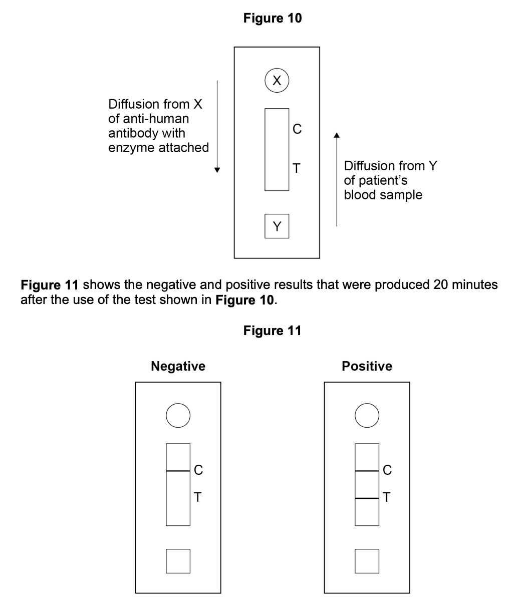
Dengue fever is a human disease caused by the dengue virus. Scientists designed an ELISA test to detect antibodies to the dengue virus in a patient’s blood sample. Figure 10 shows a diagram of this test and some information about how it works.
Suggest what is on the test at line T and explain what causes the line to appear in a positive test.
Hint
Read the question carefully: what is being detected, antibodies or antigens? Describe how the ELISA technique produces a visible result.
Mark Scheme
- Antigen (at T) and substrate (1 mark)
- Enzyme-substrate complex (produces a line/colour change) OR Enzyme (binds) with substrate (produces line/colour change) (1 mark)
Comments from mark scheme
- Reject “antigen in blood”
- Accept “colourless dye” for substrate
- Accept “ES complex” in this instance
Tips from examiner reports
- In an ELISA-based test, the test detects antibodies (not viruses or antigens) in the blood sample
- Read the question carefully to identify exactly what the test is detecting
- Include the full sequence: antibody binds → enzyme catalyses reaction with substrate → colour change
Comments from mark scheme