DNA probes & hybridisation
What you need to know (based on the AQA specification)
What you need to know (based on the AQA specification)
The use of labelled DNA probes and DNA hybridisation to locate specific alleles of genes.
- DNA probes and DNA hybridisation allow us to diagnose genetic diseases by helping us to identify specific disease-causing alleles in DNA sequences.
What are DNA probes & hybridisation?
-
DNA probe = Short single-stranded length of DNA with a label, which makes it identifiable. Examples of DNA probes:
- Radioactively labelled probes
- Detected using X-ray film or autoradiography
- Fluorescently labelled probes
- Emit light when exposed to UV light
- Radioactively labelled probes
-
DNA hybridisation = The process where a single-stranded DNA probe binds to a complementary single-stranded DNA sequence.
- DNA hybridisation: checking for a mutant allele
- Identify the DNA sequence of the mutant allele. The base sequence of the harmful mutation is known from DNA sequencing.
- Produce a DNA probe. A short single-stranded DNA sequence with complementary bases to the mutant allele is made and a fluorescent label is attached.
- Obtain DNA from the person being tested. DNA is extracted from cells (e.g. saliva).
- Amplify the person’s DNA if necessary (PCR). PCR is used to produce enough copies of the target DNA region.
- Heat is used to separate the person’s double-stranded DNA into single strands.
- Mix the DNA probes with the person’s single-stranded DNA.
- DNA hybridisation occurs if the mutant allele is present. The probe binds to the complementary DNA sequence by base pairing.
- Detect the probe. Fluorescence is detected under UV light, indicating the presence of the mutant allele.

In addition to the process above, we can use microarrays which allows the screening of multiple genes using different DNA probes. This saves the person having to do multiple different screenings for different genes.
DNA Probe & Hybridisation Applications
What you need to know (based on the AQA specification)
What you need to know (based on the AQA specification)
The use of labelled DNA probes that can be used to screen patients for heritable conditions, drug responses or health risks. The use of this information in genetic counselling and personalised medicine.
Genetic Screening
- DNA probes and DNA hybridisation can be used in genetic screening to identify disease-causing alleles in an individual’s DNA. Genetic screening has many uses, including:
- Detecting whether an individual is at a higher risk of developing certain genetic diseases caused by known mutations, for example:
- Cystic fibrosis (mutations in the CFTR gene)
- Huntington’s disease (expanded repeat in the HTT gene)
- Detecting whether an individual is a carrier of a recessive genetic disease
- Carriers do not show symptoms but can pass the allele to their offspring.
- Detecting oncogenes or mutated tumour-suppressor genes, allowing identification of individuals at a higher risk of developing certain cancers, for example:
- BRCA1 and BRCA2 mutations (increased risk of breast and ovarian cancer)
Genetic Counselling
The results from this genetic screening can be used in genetic counselling. This involves providing advice to help individuals:
- understand their risk of inheriting or passing on genetic conditions
- make informed decisions about family planning and lifestyle changes (e.g alterations in diet)
Personalised Medicine
The results from genetic screening can also be used in personalised medicine. Personalised medicine involves tailoring medical treatment to an individual based on their DNA.
This allows doctors to:
- select the most effective drug and appropriate dosage for individuals
- avoid drugs that are less effective due to genetic variation
- reduce the risk of adverse drug reactions
Exam Question Practice
What is a DNA probe?
(2 marks)Mark Scheme
- A DNA probe is a short, single-stranded piece of DNA with a sequence complementary to the target DNA (1 mark)
- It carries a fluorescent or radioactive label so that when it hybridises (binds), it can be detected (1 mark)
DNA probes are used to detect specific base sequences of DNA. The process is shown in Figure 6.
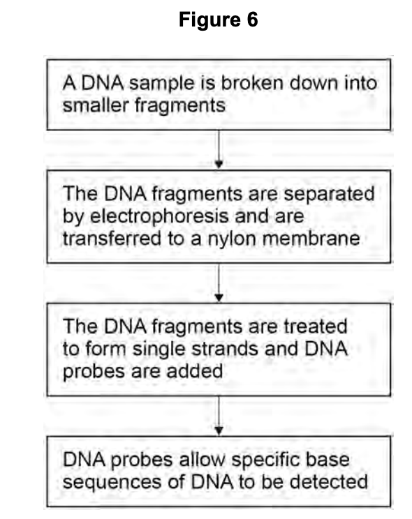
Describe how the DNA is broken down into smaller fragments.
(2 marks)Mark Scheme
- The DNA is cut using a restriction endonuclease (restriction enzyme) (1 mark)
- This enzyme recognises and cuts at a specific base sequence (recognition/restriction site), breaking the phosphodiester bonds (1 mark)
The DNA on the nylon membrane is treated to form single strands. Explain why.
(1 marks)Hint
Think about how DNA probes bind to their target sequence.
Mark Scheme
- The DNA must be single-stranded so that the DNA probe can hybridise (bind by complementary base pairing) to its target sequence (1 mark)
The scientists used a radioactively labelled DNA probe to show that the cells of tobacco plant leaves contained the SUT1 gene. Describe how they would do this. Do not include PCR in your answer.
(4 marks)Mark Scheme
- Extract DNA from the leaf cells and cut it into fragments using restriction endonucleases (1 mark)
- Separate the DNA fragments by size using gel electrophoresis (1 mark)
- Denature the DNA (e.g. by heating) to produce single-stranded DNA — this exposes the bases so the probe can bind (1 mark)
- Add the radioactively labelled DNA probe, which hybridises (binds by complementary base pairing) to the SUT1 gene sequence (1 mark)
- Use autoradiography (X-ray film) to detect where the radioactive probe has bound, revealing the location of the SUT1 gene (1 mark)